

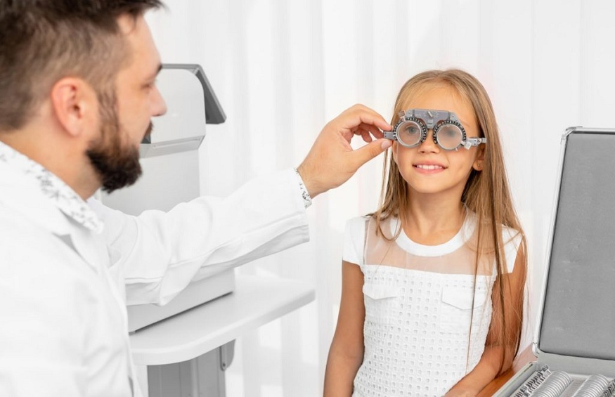
Properly evaluating pupils is critical for neurological and eye assessments. Subtle pupil changes can provide valuable diagnostic clues, making accurate measurement vital. However, inconsistencies occur with manual methods.
This is where the NPi (Neurological Pupil index) is transformative. The NPi utilizes advanced pupillometry to enable unprecedented precision in measuring pupil characteristics. Let’s explore how this innovative technology changes pupil measurement and enhances medical exams.
Understanding Pupil Measurement Significance
During neurological evaluations, clinicians routinely assess pupil size and reactivity. Changes can signify dysfunction from stroke, increased intracranial pressure, optic nerve damage, and more. For instance, pupils demonstrating sluggish reactions to light or asymmetric sizes may indicate underlying neuropathology necessitating further assessment.
Precisely measuring pupils is key for detecting small variations that provide diagnostic insights. However, studies show inconsistent agreement between practitioners using manual neurological tools. Thus, more rigorous, objective pupillary measurements are needed to optimize diagnoses. This is the need that NPi technology strives to meet.
Introducing the NPi
The NPi, short for Neurological Pupil Index, represents a major advance in objective pupillary measurement. This innovative pupillometry system utilizes infrared cameras and specialized algorithms to accurately measure pupil size, percentage of constriction, and light reaction latency.
The NPi generates a score from 0 to 5, quantifying pupil reactivity. This provides an objective reference metric unaffected by issues like ambient lighting that introduce variability in manual evaluations. NPi enables enhanced accuracy, consistency, and standardization of pupil measurements.
The Science Behind NPi
The foundation of the NPi system is the proprietary algorithm for calculating pupillary reactivity metrics. It analyzes high-resolution infrared video of the pupil’s responses to calibrated light pulses. Frame-by-frame changes in pupil diameter are measured, and reaction latencies are determined.
Importantly, the algorithm accounts for ambient lighting, patient positioning, and anatomy variations. It computes a normalized NPi score by comparing stimulated and non-stimulated pupil characteristics. This makes the NPi score highly consistent across different evaluation settings and individuals.
NPi Advantages Over Traditional Methods
NPi offers significant advantages compared to conventional manual pupil measurement techniques. Traditional approaches rely on qualitative visual inspection and manual documentation of findings. This introduces considerable variability based on ambient lighting conditions, individual examiner proficiency, and intrinsic human observational error. Subtle pupillary changes often go undetected.
In contrast, NPi provides precisely measured, objective pupillary metrics using advanced infrared sensors and algorithms. It is unaffected by lighting conditions or the subjectivity of the operator. NPi enables rapid automated measurements within seconds, eliminating the need for slow, error-prone manual assessments. The sensitive infrared video capture can discern minute pupillary changes that are impossible to visualize with the naked eye. This allows the identification of subtle neurological abnormalities.
NPi and Individualized Care
A major NPi benefit is that it enables physicians to develop treatment plans based on objective pupillary data. The pupillary light reflex pathway includes the retina, optic nerve, and midbrain oculomotor nucleus.
Having precise NPi measurements and pupillary reaction waveforms allows for pinpointing areas of dysfunction along this pathway. This assists in crafting targeted treatment strategies tailored to each patient’s particular neurological deficits evident on their pupillary exam.
Streamlining Documentation and Analysis
NPi helps to automate data collection, documentation, and analysis. Pupil measurements are instantly recorded electronically for seamless integration into digital health platforms.
Clinicians can efficiently track longitudinal pupillary trends using sequential NPi scores. And pupillary reaction waveform patterns can be compared side-by-side. This streamlines record-keeping and enhances retrospective review capabilities.
Incorporating NPi into Clinical Workflows
Integrating NPi pupillometry into busy practice workflows is readily achievable. The portable system is designed for ease of use. With brief training, nurses can capture high-quality pupil measurements in about 30–60 seconds per patient.
Clinicians simply review automated NPi scores and waveforms to interpret results. Testing can occur at intake alongside standard vitals. Periodic assessments enable tracking changes. With minimal practice, NPi blends seamlessly into existing clinical evaluation routines, strengthening assessments.
Future Implications of NPi Technology
While currently focused on pupil measurement, the core NPi technology shows promise for even broader applications utilizing iris pattern analysis for medical diagnostics and tracking neurological status over time.
Ongoing research aims to expand NPi capabilities. One example is using NPi video to non-invasively detect intracranial pressure. As the algorithms, camera resolution, and computing power improve, NPi is poised to become an increasingly versatile neuro exam metric.
Conclusion:
The NPi allows unprecedented consistency and accuracy in pupil measurement. This enables clinicians to gain superior diagnostic insights, inform individualized treatments, and streamline neurological evaluations. By embracing advanced technologies like NPi, medical professionals can continue elevating patient care standards through enhanced precision.